

Peripheral angiography is a test similar to coronary angiography. Unlike coronary angiography which is used to look at the condition of the artery supplying your heart, peripheral angiography is used to check the condition of blood vessels supplying your legs and arms. This test uses a special x-ray machine and it is performed in a cath lab.
When To Do Peripheral Angiography?
A peripheral angiogram is useful to test for the diagnosis of peripheral artery disease (PAD). PAD is a disease in which cholesterol and/or blood clots cause narrowing of the lumen of the artery supplying blood to the legs or arms. Such blocks in the artery cause muscle pain or cramping in legs or arms on activity, such as walking or climbing stairs and disappears after a few minutes of rest (intermittent claudication).
Once the disease progresses, you may feel pain even at rest. Critical narrowing of the artery can cause ulcer or gangrene formation in your leg.
Peripheral Angioplasty
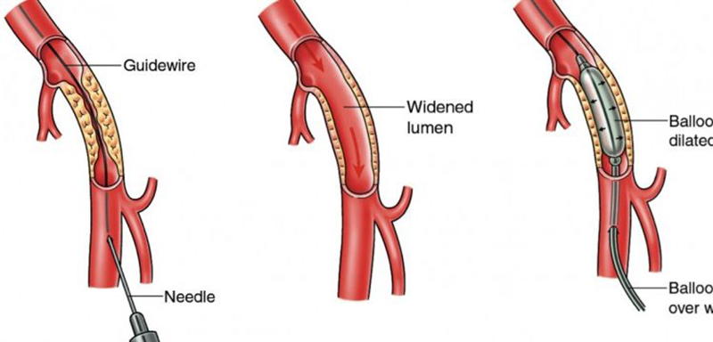
Treatment for peripheral artery disease includes special drugs that reduce the pain in your leg while walking or climbing stairs. If symptoms are not relieved with the medications then angioplasty or surgery is indicated. In peripheral angioplasty, a catheter (thin hollow tube) is inserted through your groin. Then in this catheter, a balloon is advanced in the blocked artery.
The balloon is then inflated; this widens the artery and reduces the block. A stent (a small mesh tube) may be placed in the artery during angioplasty. A stent helps keep the artery open and prevent re-occlusion.
Catheter-Directed Thrombolysis (CDT)
Sometimes your artery may be blocked due to a blood clot in it. In such situation injection of a clot-dissolving drug (clot busters) into your artery through a catheter (thin hollow tube) is done to break the clot and to restore the blood flow in the blocked artery.